Check out this answer from Consensus:
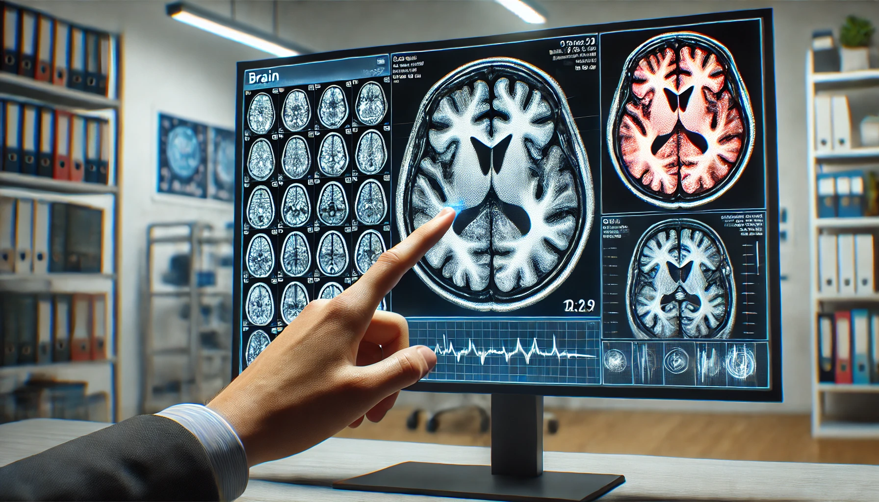
Brain imaging can indeed detect anxiety by identifying specific patterns of brain activity and structural abnormalities associated with different anxiety disorders. Key regions such as the amygdala, anterior cingulate cortex, and insula play crucial roles in the manifestation of anxiety. Functional and structural neuroimaging studies provide consistent evidence of these neural correlates, supporting the use of brain imaging as a tool for understanding and potentially diagnosing anxiety disorders.
Anxiety disorders are a group of mental health conditions characterized by excessive fear and anxiety. Recent advancements in brain imaging techniques have provided valuable insights into the neurobiological underpinnings of these disorders. This synthesis aims to determine whether anxiety can be detected through brain imaging by analyzing findings from multiple research papers.
Key Insights
- Role of Specific Brain Regions:
- The amygdala, anterior cingulate cortex, and insula are consistently implicated in the pathophysiology of various anxiety disorders, showing hyperactivation in response to anxiety-provoking stimuli1 2 3 4 5.
- Structural abnormalities in the hippocampus and orbitofrontal cortex are also noted, particularly in posttraumatic stress disorder (PTSD) and obsessive-compulsive disorder (OCD)5 6.
- Functional Neuroimaging Findings:
- Functional MRI (fMRI) studies reveal increased activity in limbic and paralimbic regions, especially the amygdala, in social anxiety disorder (SAD) and specific phobia2 3.
- PTSD patients show hypoactivation in the dorsal and rostral anterior cingulate cortices and the ventromedial prefrontal cortex, which are linked to emotional regulation2.
- Transdiagnostic and Disorder-Specific Patterns:
- Both major depressive disorder (MDD) and anxiety disorders share alterations in the orbitofrontal cortex, middle frontal cortex, and limbic regions, suggesting common neural mechanisms4.
- Anxiety-specific functional alterations are observed in the insula and frontal regions during emotional tasks, and in the inferior parietal lobule and superior frontal gyrus during cognitive tasks4.
- Trait and State Anxiety:
- Resting-state fMRI studies indicate that intrinsic brain activity in the right ventral medial prefrontal cortex and dorsal anterior cingulate cortex is associated with trait anxiety. Changes in the right insula’s activity predict state anxiety variability9.
Can anxiety be detected from brain imaging?
Laura Schrader has answered Uncertain
An expert from Tulane University in Molecular Biology, Neurobiology, Neuroimaging, Neuroscience
Anxiety itself can not be detected by brain imaging, but aberrant activity of brain areas associated with anxiety, such as the amygdala, can be measured by brain imaging.
Can anxiety be detected from brain imaging?
Milena de Barros Viana has answered Likely
An expert from Federal University of São Paulo in Neuropsychology
By using brain imaging it is possible to see that some structures are more activated in patients with anxiety related-disorders (for instance the amygdala in generalized anxiety) and the periaqueductal grey (in panic disorder patients).
