Will an MRI Affect Dental Fillings?

Have a question about science, health, fitness, or diet? Get cited, evidence-based insights: Consensus is an AI-Powered Academic Search Engine.

Try for free
Contents
    Written by Consensus AI
    5 min read

    Check out this answer from Consensus:

    The interaction between MRI and dental fillings, particularly those containing mercury, is a complex issue that warrants careful consideration. Research indicates that high-field MRI can increase mercury release from dental amalgam fillings, posing potential health risks. Additionally, dental fillings can cause artifacts in MR images, affecting diagnostic accuracy. As MRI technology continues to evolve, further studies are needed to develop safer imaging protocols and improve our understanding of the effects of MRI on dental materials.

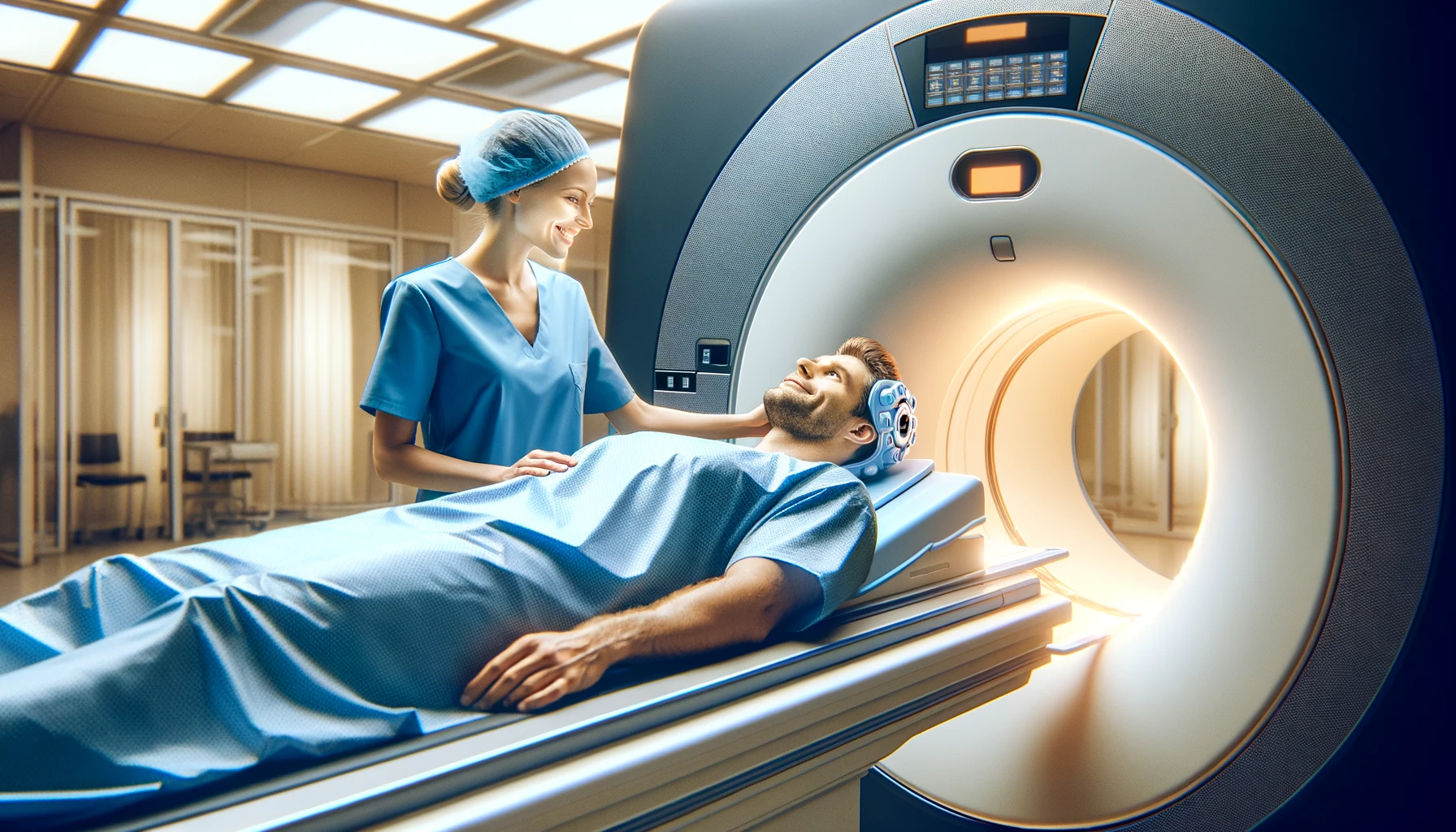
    Magnetic Resonance Imaging (MRI) is a widely used diagnostic tool in modern medicine, known for its ability to produce detailed images of the body’s internal structures without the use of ionizing radiation. However, concerns have been raised about the potential effects of MRI on dental fillings, particularly those containing metals such as mercury. This article explores the impact of MRI on dental fillings, drawing on findings from recent research studies.

    Dental amalgam fillings, which contain a mixture of metals including mercury, have been used for over a century to restore decayed teeth. Despite their durability and cost-effectiveness, the safety of amalgam fillings has been a topic of debate due to the potential release of mercury, a toxic element. With the increasing use of high-field MRI in medical diagnostics, it is crucial to understand whether exposure to MRI can exacerbate mercury release from dental fillings and pose a health risk to patients.

    Mercury Release from Dental Amalgam Fillings

    High-Field MRI and Mercury Release

    A study investigated the effect of high-field MRI (1.5 Tesla) on mercury release from dental amalgam fillings. The study involved two groups of students with similar dental restorations, one group exposed to MRI and the other serving as a control. The results showed a significant increase in urinary mercury levels in the MRI-exposed group compared to the control group, indicating that MRI exposure can indeed enhance mercury release from dental amalgam fillings.

    Ex Vivo Studies on Mercury Release

    Another study examined mercury release from dental amalgam fillings in an ex vivo setting, using both 1.5-T and 7.0-T MRI. The findings suggested that higher magnetic field strengths could potentially trigger greater mercury release. This study highlights the need for further research to fully understand the implications of high-field MRI on dental amalgam fillings.

    MRI Artifacts and Dental Fillings

    Artefacts in MR Imaging

    Dental fillings, particularly those containing metals, can cause artifacts in MR imaging. An investigation into various dental filling materials revealed that while most materials did not cause significant artifacts, one type did produce noticeable distortions in the images. This suggests that the composition of the filling material plays a crucial role in the extent of MRI artifacts.

    Advanced Imaging Techniques

    Recent advancements in MRI technology, such as ultra-short echo time (UTE) MRI, have shown promise in reducing artifacts and improving the assessment of dental hard tissues. These techniques allow for better visualization of dental structures and may mitigate some of the issues associated with traditional MRI imaging of dental fillings.

    Clinical Implications

    Safety and Diagnostic Accuracy

    The potential release of mercury from dental amalgam fillings during MRI procedures raises important safety concerns. Patients with amalgam fillings should be informed about the possible risks, and alternative imaging methods may be considered for those with a high number of amalgam restorations. Additionally, the presence of dental fillings can affect the diagnostic accuracy of MRI scans due to artifacts, which may necessitate the use of advanced imaging techniques or alternative diagnostic tools .

    Recommendations for Dental Practice

    Given the findings from recent studies, dental practitioners should be aware of the potential interactions between MRI and dental fillings. It is advisable to conduct a thorough assessment of a patient’s dental restorations before recommending MRI procedures. In cases where MRI is necessary, informing the radiologist about the presence of dental fillings can help in planning the imaging protocol to minimize artifacts and ensure accurate diagnosis.


    Will an MRI affect dental fillings?

    Rebecca Dewey has answered Unlikely

    An expert from The University of Nottingham in Neuroimaging, Neuroscience

    It’s unlikely to – your fillings don’t contain magnetic materials and they don’t form a current loop, i.e. a circuit through which an electrical current can flow. You might experience a metallic taste in your mouth but that’s not the scan per se – it’s the magnetic field gradient (i.e. the magnetic field changing over time or you moving from one area of the field to another) acting on your saliva/taste buds. That taste will stop as soon as you stay still or leave the field.

    What’s more interesting though is the effect of your fillings on the MRI scan. The metal won’t generate any signal as it contains no water, and it will also cause there to be no signal in the surrounding areas, so you will look like those teeth are missing! No harm done though and your teeth are far enough away from your brain that it won’t make your images any less useful!

    Have a question about science, health, fitness, or diet? Get cited, evidence-based insights: Consensus is an AI-Powered Academic Search Engine.

    Try for free
    Contents